
Fig. 3
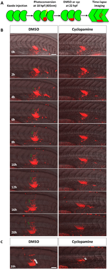
Lineage tracing of sclerotome derived cells. (A) Schematic of the experimental procedure. Embryos were injected with Kaede mRNA at the one-cell stage. At 18 hpf, Kaedegreen in the presumptive ventral sclerotome of a newly formed somite (somite 17 or 18) was photoconverted to Kaedered. At 22 hpf, photoconverted embryos were treated with either DMSO or cyclopamine and imaged over the next 20 hours. (B) Representative snapshots of different time-points during lineage tracing of sclerotome derived cells between 22 hpf (0h) and 42 hpf (20h). Merged images containing the red channel and the bright field are shown. Kaedered sclerotome derived cells in DMSO treated controls migrated from the ventral sclerotome to the notochord before dividing to generate a population of cells surrounding the notochord. Green arrows indicate one representative sclerotome derived cell and its progeny. In the presence of cyclopamine, sclerotome derived cells failed to migrate out of the ventral sclerotome to the notochord. The corresponding movies are shown in S1 and S2 Videos. n = 7 embryos per condition. (C) Projection of deep confocal slices at the last time point of time-lapse. A population of Kaedered cells reminiscent of tenocytes (arrowheads) located along the myotendinous junction between somites (dotted lines). Fewer tenocyte-like cells were found in cyclopamine treated embryos. Scale bars: 50 μm. |