
Fig. 3
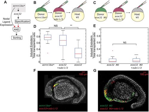
The combination of Nodal ligand expression and endodermal fate is sufficient to trigger ectopic endodermal cell sorting. (A) Schematic depicting putative AND gate for endoderm sorting. Red arrows demonstrate the experimental perturbation to test the sufficiency of Nodal ligands to induce ingression. (B,C) Schematics of double transplantation assay to test the sufficiency of the AND gate depicted in A for endodermal sorting. Cells overexpressing acvr1ba* (green) were transplanted together with cells overexpressing sox32, ndr1 and ndr2 (red) into the animal pole of a wild-type (WT) host embryo (B). Cells containing sox32 MO only were transplanted together with cells containing sox32 MO as well as ndr1 and ndr2 mRNAs were transplanted into the animal pole of a wild-type host embryo (C). (D) Boxplot quantification of endoderm contribution at 20 hpf of the transplanted cells depicted in B. Cells overexpressing sox32, ndr1 and ndr2 contributed to endoderm at a similar rate compared with cells overexpressing acvr1ba*. Data are shown as mean±s.e.m. of three independent transplantation experiments with 18 embryos per condition. **P<0.01 (Student's t-test). (E) Boxplot quantification of endoderm contribution at 20 hpf of the transplanted cells depicted in C. Neither cells containing sox32 MO nor cells containing sox32 MO and overexpressing ndr1 and ndr2 contributed to endodermal tissue. In addition, cells expressing acvr1ba* and sox32 MO did not contribute to endodermal tissue. Data are shown as mean±s.e.m. of two independent transplantation experiments with 14 embryos per condition. Student's t-test. (F) Representative image showing distribution of the transplanted cells depicted in D at the 18-somite stage. acvr1ba*-expressing cells localize to the endoderm-derived tissue, primarily the pharynx (green). Cells overexpressing sox32, ndr1 and ndr2 also localize to endoderm-derived tissue, primarily the pharynx (red). Lateral view, anterior to the left. (G) Representative image showing distribution of the transplanted cells depicted in E at the 18-somite stage. Cells expressing sox32 and Nodal ligands (ndr1, ndr1) localize to endodermal tissues similar to cells expressing acvr1ba*. In contrast, sox32 MO-injected cells (green) and cells injected with sox32 MO and ndr1 and ndr2 mRNAs (red) localize to non-endodermal tissue, primarily in the head and skin. Lateral view, anterior to the left. NS, not significant. |