
Fig. 5
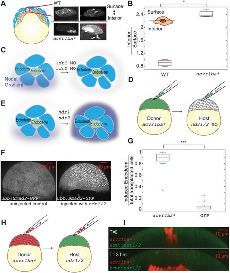
Nodal ligands initiate but do not guide the ingression of endodermal cells. (A) Actin localization in ectopic endodermal cells. Blue, ectoderm; brown, mesoderm; green, endoderm. Donor embryos were injected with GFP-UTRN mRNA to label actin filaments. Cells overexpressing acvr1ba* or control cells expressing GFP-UTRN only were transplanted to the animal poles of wild-type host embryos. Actin was enriched on the interior side of acvr1ba*-expressing cells whereas control cells exhibited uniform actin distribution. Data were re-sliced and projected to the xz plane, with the surface of the embryo towards the top and the interior towards the bottom. Arrow shows interior-facing protrusion. (B) Boxplot of the ratio of interior to surface accumulation of actin. acvr1ba*-expressing cells exhibited significant interior enrichment of actin compared with control cells. Data are shown as mean±s.e.m. of three independent transplantation experiments, with 58 cells per condition. *P<0.05 (Student's t-test). Schematic shows the segmentation of surface versus interior of an endodermal cell. (C,E) Investigation into whether the endogenous Nodal gradient functions as a directional cue for endoderm ingression through knockdown of endogenous Nodal ligands. (C) ndr1 and ndr2 MOs were injected into host embryos to remove the endogenous Nodal gradient. (D) Cells expressing acvr1ba* were transplanted to the animal pole of a Nodal-depleted host. (G) Boxplot quantification of endoderm contribution at 20 hpf for all transplanted cells. Cells overexpressing acvr1ba* still contributed to endodermal tissues even in the absence of an endogenous Nodal gradient. Data are shown as mean±s.e.m. of two independent transplantation experiments, with 15 embryos per condition. ***P<0.001 (Student's t-test). (E) Saturating the endogenous Nodal gradient to test whether it acts as a directional cue. (F) Tg(ubb:Smad2-GFP) shows uniform nuclear translocation in a ndr1 and ndr2-injected embryo, suggesting uniform Nodal signaling. (H) Cells expressing acvr1ba* were transplanted to the animal pole of a Nodal-saturated host. (I) Representative image showing positions of acvr1ba* cells (red) immediately and 3 h after transplantation in a Nodal-saturated host. |