
Fig. 2
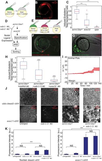
Nodal ligand expression is necessary to trigger the sorting of ectopic acvr1ba*-induced endodermal cells. (A,B) Schematics depicting sox32-induced ectopic endoderm transplant assay (A) and representative result (B). sox32-overexpressing cells were transplanted to the animal pole of wild-type host embryos. At the 21-somite stage, transplanted sox32-overexpressing cells primarily localized to non-endodermal tissues, primarily in the head and skin. (C) Boxplot quantification of endoderm contribution of transplanted cells at 20 hpf, assessed by colocalization with Tg(sox17:dsRed) expression. Here and in subsequent figures, for each box, the central mark indicates the median, and the bottom and top edges of the box indicate the 25th and 75th percentiles, respectively. The whiskers extend to the most extreme data points not considered outliers, and the outliers are plotted individually using the ‘+’ symbol. Compared with acvr1ba*-expressing cells, fewer cells overexpressing sox32 contributed to endodermal tissues. Data are shown as mean±s.e.m. of three independent transplantation experiments with 26 embryos per condition. ***P<0.001 (Student's t-test). (D) Schematic depicting a potential AND gate for endoderm sorting. Constitutively activate acvr1ba* upregulates both sox32 as well as Nodal ligand expression. Only with both inputs do cells successfully sort to the inner layer of the embryo. (E) Schematic of the cell transplantation assay to test the necessity of Nodal ligand expression for cell sorting. Donor cells containing ndr1 and ndr2 MOs plus acvr1ba* mRNA (red) were transplanted together with cells overexpressing acvr1ba* only (green) into the animal pole of a wild-type (WT) host embryo. (F) Representative images showing the distribution of transplanted cells at the 21-somite stage. Cells expressing acvr1ba* only (green) localize to endoderm-derived tissue, primarily the pharynx. Cells containing acvr1ba* along with ndr1 MO and ndr2 MO (red) localize to non-endodermal tissue, primarily in the head. Lateral view, anterior to the bottom-left. (G) Representative image showing the distribution of transplanted cells at the 21-somite stage. Cells expressing acvr1ba* along with sox32 MO (green) localize to ectoderm-derived tissue, primarily the head. Lateral view, anterior to the bottom-left. (H) Boxplot quantification of endoderm contribution of transplanted cells at 20 hpf. ndr1 and ndr2 knockdown as well as sox32 knockdown reduced the ability of acvr1ba*-expressing cells to contribute to endodermal tissue. Data are shown as mean±s.e.m. of three independent transplantation experiments with 22 embryos per condition. ***P<0.001 (Student's t-test). (I) Single-cell tracking analysis of ingression of acvr1ba*-expressing cells with ndr1 MO and ndr2 MO. Average relative distance (±s.e.m.) plotted against time. Relative distance was calculated as in Fig. 1H. (J) Nodal signaling levels assessed by Tg(ubb:Smad2-GFP) and Tg(sox17:GFP). Top: Smad2-GFP showed no nuclear localization in cells at the animal poles (AP) of uninjected embryos and ndr1/2 morphants. Smad2-GFP showed comparable levels of nuclear localization in acvr1ba*-injected embryos and acvr1ba* with ndr1 and ndr2 MO-injected embryos. Bottom: Sox17:GFP labels wild-type endodermal cells in the uninjected control embryo but few GFP-positive cells are present in the ndr1/2 morphants. Sox17:GFP shows elevated level of expression in both acvr1ba*-injected embryos and acvr1ba* with ndr1 and ndr2 MO-injected embryos. Animal pole view. (K) Quantification of Nodal signaling level. Nuclear Smad2-GFP and Sox17:GFP fluorescence levels are quantified. Data are shown as mean±s.e.m. of three independent embryos. ***P<0.001 (Student's t-test). NS, not significant. |