
Fig. 4
- ID
- ZDB-FIG-180914-37
- Publication
- Cantaut-Belarif et al., 2018 - The Reissner Fiber in the Cerebrospinal Fluid Controls Morphogenesis of the Body Axis
- Other Figures
- All Figure Page
- Back to All Figure Page
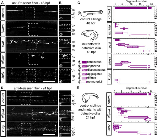
Intact Cilia Are Necessary for the Formation of the Reissner Fiber in the Central Canal (A) Z projection of stacks of lateral optical sections (depth 4–5 μm) of spinal cord at 48 hpf showing modifications of immunoreactivity for the Reissner fiber in mutants with defective cilia (iguana, oval, elipsa, and kurly) compared to control siblings, where a continuous fiber runs along the entire central canal. d, dorsal; v, ventral. Scale bar represents 30 μm. (B) Zoom of regions boxed in (A) showing depositions of the Reissner material in the central canal. Deposits occur as continuous densely packed fiber (1), continuous unpacked fiber (2), discontinuous loosely packed material (3), aggregated material (4), diffuse material (5), and absence of material (6). cc, central canal; fp, floor plate; nc, notochord. (C) Distribution of defects in the Reissner material along the rostro-caudal axis and for each mutant displayed as mean segment number ± SEM (n = 24; 11; 6; 8; 5 embryos for control siblings; iguana; oval; elipsa, and kurly, respectively). (D and E) Before elipsa and kurly embryos develop the curled-down phenotype (24 hpf embryo), defects in the Reissner material can be observed in the central canal. Right panels (D) show zoom from regions boxed in left panels with depositions of the Reissner material in the central canal; the numbers correspond to the equivalent defects at 48 hpf (depicted in B and C). Distribution of defects in the Reissner material along the rostro-caudal axis for the two mutants at 24 hpf (E) (bottom, n = 12 and 19 embryos for elipsa and kurly, respectively) compared to control siblings (top; n = 28). Scale bar represents 30 μm. Cc, central canal; fp, floor plate; nc, notochord. See also Figure S4. |
| Antibody: | |
|---|---|
| Fish: | |
| Anatomical Term: | |
| Stage Range: | Prim-5 to Long-pec |
| Fish: | |
|---|---|
| Observed In: | |
| Stage Range: | Prim-5 to Long-pec |