
Fig. 2
- ID
- ZDB-FIG-180914-35
- Publication
- Cantaut-Belarif et al., 2018 - The Reissner Fiber in the Cerebrospinal Fluid Controls Morphogenesis of the Body Axis
- Other Figures
- All Figure Page
- Back to All Figure Page
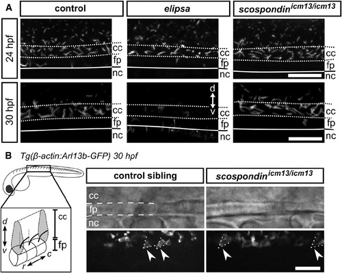
Structural and Dynamic Properties of Cilia Appear Intact in the scospondinicm13/icm13 Mutant (A) Z projection of stacks of lateral optical sections (depth 4–5 μm) of spinal cord immunostained against acetylated tubulin show intact cilia projecting into the central canal of control and scospondinicm13/icm13 larvae at 24 (top) and 30 hpf (bottom). In comparison, elipsa embryos exhibit fewer cilia at 24 hpf, which are not maintained at 30 hpf. Scale bars represent 15 μm. (B) Time projection from a 30-s-long time series acquired at 17 Hz and indicating movement of cilia expressing GFP in Tg(β-actin:Arl13b-GFP; scospondinicm13/icm13) animals (right) and control siblings (left). Note the similarity in position (posterior tilt, dashed lines) and beating amplitude of motile cilia in the central canal (arrowheads) in mutant embryos compared to control siblings. The schematic summarizes our observations. Rostral, left; dorsal, top. Scale bar represents 10 μm. See also Figure S2 and Video S1. |
| Gene: | |
|---|---|
| Antibody: | |
| Fish: | |
| Anatomical Terms: | |
| Stage Range: | Prim-5 to Prim-15 |
| Fish: | |
|---|---|
| Observed In: | |
| Stage Range: | Prim-5 to Prim-15 |