
Fig. S4
- ID
- ZDB-FIG-180914-41
- Publication
- Cantaut-Belarif et al., 2018 - The Reissner Fiber in the Cerebrospinal Fluid Controls Morphogenesis of the Body Axis
- Other Figures
- All Figure Page
- Back to All Figure Page
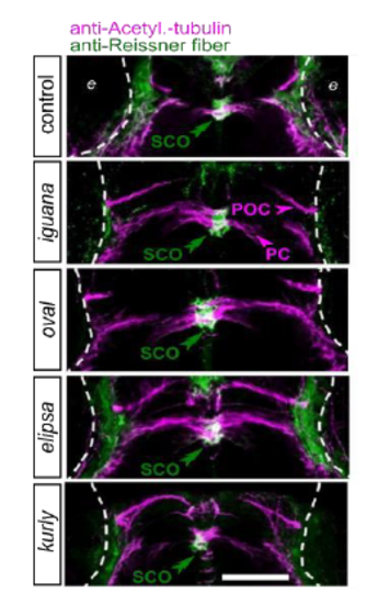
The sub-commissural organ is immunoreactive for the Reissner fiber material in mutants with defective cilia, related to Figure 4. Z projection of stacks of dorsal optical sections (depth = 23 - 26 μm) of 48 hpf embryos immunostained against Acetylated-tubulin (magenta) allowing to detect axonal tracts (POC: post-optic commissure, PC: posterior commissure, arrowheads), and the Reissner fiber material (green) in the sub-commissural organ (SCO, double arrowheads). Control embryos (here an iguana sibling) as well as iguana, oval, elipsa and kurly mutants show immunoreactivity for the Reissner fiber material in the SCO. Rostral, top. e: eye. Scale bar represents 50 μm. |