
Fig. S3
- ID
- ZDB-FIG-180914-40
- Publication
- Cantaut-Belarif et al., 2018 - The Reissner Fiber in the Cerebrospinal Fluid Controls Morphogenesis of the Body Axis
- Other Figures
- All Figure Page
- Back to All Figure Page
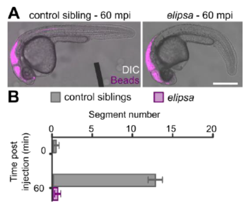
Transport of exogenous fluorescent beads in the cerebrospinal fluid is abolished in elipsa mutants with defective cilia, related to Figure 3 and Video S2. A. Superimposed images of transmitted light (DIC) and fluorescent 20-nm diameter beads (magenta) injected in the third ventricle of 30 hpf control sibling and elipsa mutant embryos. Beads are transported down the central canal in straight control siblings, but not in elipsa mutants as shown 60 minutes post-injection (mpi). Scale bar represents 0.5 mm. The progression of the fluorescence front in the central canal is quantified in B as the segment number reached 60 minutes after injection (mean ± SEM) in control sibling (n = 3) and elipsa mutant embryos (n = 5) (p = 0.0014; t = 12.46, df = 2.84, two tailed t-test). |