
FIGURE
Fig. S6
- ID
- ZDB-FIG-180912-41
- Publication
- Morris et al., 2018 - Live imaging of collagen deposition during skin development and repair in a collagen I - GFP fusion transgenic zebrafish line
- Other Figures
- All Figure Page
- Back to All Figure Page
Fig. S6
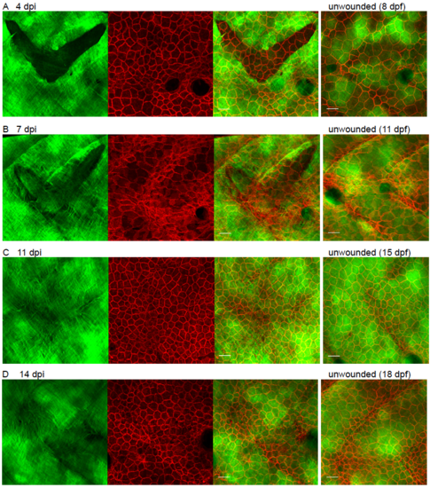
Migration of epidermal cells versus epidermal collagen I deposition post wounding. Still images from repairing wounds to the flanks of 4 dpf Tg(krt19:col1a2-GFP), Tg(krt19:tdTomatoCAAX) double transgenic fish to reveal relative time course of repair of the cellular versus matrix layers. Scale bar = 25 μm. |
Expression Data
Expression Detail
Antibody Labeling
Phenotype Data
Phenotype Detail
Acknowledgments
This image is the copyrighted work of the attributed author or publisher, and
ZFIN has permission only to display this image to its users.
Additional permissions should be obtained from the applicable author or publisher of the image.
Reprinted from Developmental Biology, 441(1), Morris, J.L., Cross, S.J., Lu, Y., Kadler, K.E., Lu, Y., Dallas, S.L., Martin, P., Live imaging of collagen deposition during skin development and repair in a collagen I - GFP fusion transgenic zebrafish line, 4-11, Copyright (2018) with permission from Elsevier. Full text @ Dev. Biol.