
Fig. 2
- ID
- ZDB-FIG-180912-35
- Publication
- Morris et al., 2018 - Live imaging of collagen deposition during skin development and repair in a collagen I - GFP fusion transgenic zebrafish line
- Other Figures
- All Figure Page
- Back to All Figure Page
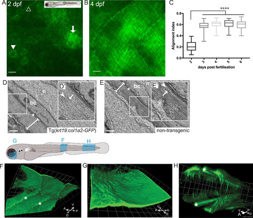
Imaging the deposition of collagen beneath the embryonic epidermis. (A) In the region of flank indicated by the box in larval schematic, the earliest GFP-collagen I is seen at 2 dpf in maximum projection confocal images of Tg(krt19:col1a2-GFP) transgenic zebrafish; GFP-collagen is seen within sporadic cells (arrowhead), and in some adjacent patches exhibiting the beginning of orthogonal patterning (arrow), as well as within myosepta (open arrowhead). (B) At 4 dpf orthogonal structure is fully evident. (C) Quantification of collagen alignment index (AI) over developmental time. Plotted as mean ± SD and analysed using a one-way ANOVA, ****p < 0.0001, n = 10–16 fish. (D, E) Transmission electron microscopy (TEM) of flank skin of 5 dpf Tg(krt19:col1a2-GFP) transgenic (D) and non-transgenic (E) larvae (with high magnification inset, D′, E′) reveals the orthogonal layering of collagen I; arrows indicate collagen fibril; arrowhead indicates adjacent orthogonal layer of collagen fibrils; n, nucleus; bc, basal cell cytoplasm; asterisk, basement membrane; line denotes collagen I layer. (F) GFP-collagen I tethers extend deep into the tissue of the 3 dpf fish as anchors to the underlying tissue within the myoseptum (asterisks); (G) Similarly, collagenous tethers link the epidermis to deep structures in the head, as for example around the eye; and (H) bilateral developing tail tendons are seen within the posterior-most portion of the developing tail (stars). Images in F, G and H are 3D reconstructions generated using Volocity, and correspond to regions indicated in the larval schematic above. Scale bars: A,B= 15 µm; C= 0.5 µm; F,G,H 1 unit= 18.51 µm; representative image of n = 3 fish imaged. |
Reprinted from Developmental Biology, 441(1), Morris, J.L., Cross, S.J., Lu, Y., Kadler, K.E., Lu, Y., Dallas, S.L., Martin, P., Live imaging of collagen deposition during skin development and repair in a collagen I - GFP fusion transgenic zebrafish line, 4-11, Copyright (2018) with permission from Elsevier. Full text @ Dev. Biol.