
FIGURE
Fig. S3
- ID
- ZDB-FIG-180912-38
- Publication
- Morris et al., 2018 - Live imaging of collagen deposition during skin development and repair in a collagen I - GFP fusion transgenic zebrafish line
- Other Figures
- All Figure Page
- Back to All Figure Page
Fig. S3
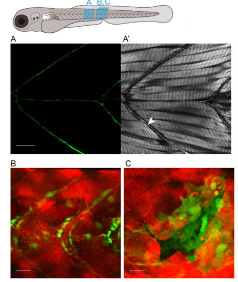
Epidermal-derived myoseptal collagen I structures and relationship of epidermal-derived mCherry-collagen I to invading ET37 fibroblast-like cells (A) Equivalent single z-plane confocal and SHG (A’) image of a 10 dpf Tg(krt19:col1a2-GFP) zebrafish, indicating myoseptal labelling (arrowhead in A’). (B,C) A maximum projection confocal images of Tg(krt19:col1a2-mcherry), ET37 double transgenic zebrafish to show relationship of epidermal-derived collagen I (red), with influx of fibroblasts-like cells (green) to wound, just prior to (B) and 2 dpi (C). Scale bars = 25 μm. |
Expression Data
Expression Detail
Antibody Labeling
Phenotype Data
Phenotype Detail
Acknowledgments
This image is the copyrighted work of the attributed author or publisher, and
ZFIN has permission only to display this image to its users.
Additional permissions should be obtained from the applicable author or publisher of the image.
Reprinted from Developmental Biology, 441(1), Morris, J.L., Cross, S.J., Lu, Y., Kadler, K.E., Lu, Y., Dallas, S.L., Martin, P., Live imaging of collagen deposition during skin development and repair in a collagen I - GFP fusion transgenic zebrafish line, 4-11, Copyright (2018) with permission from Elsevier. Full text @ Dev. Biol.