
FIGURE
Fig. S2
- ID
- ZDB-FIG-180912-37
- Publication
- Morris et al., 2018 - Live imaging of collagen deposition during skin development and repair in a collagen I - GFP fusion transgenic zebrafish line
- Other Figures
- All Figure Page
- Back to All Figure Page
Fig. S2
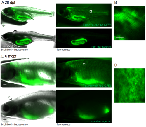
GFP expression in juvenile and adult GFP-collagen transgenic zebrafish. (A) Widefield bright field and fluorescent images of 28 dpf and (C) 6 month pf (mopf) GFP-collagen I fish showing they remain GFP positive, whereas only the gut is autofluorescent in non-transgenic fish. Region imaged by confocal indicated by white box. (B, D) Maximum projection confocal images demonstrate the GFP-collagen I is still orthogonal in nature, but it is located in the scale-layer. Scale bars: A = 1mm; B,D = 25 μm. |
Expression Data
Expression Detail
Antibody Labeling
Phenotype Data
Phenotype Detail
Acknowledgments
This image is the copyrighted work of the attributed author or publisher, and
ZFIN has permission only to display this image to its users.
Additional permissions should be obtained from the applicable author or publisher of the image.
Reprinted from Developmental Biology, 441(1), Morris, J.L., Cross, S.J., Lu, Y., Kadler, K.E., Lu, Y., Dallas, S.L., Martin, P., Live imaging of collagen deposition during skin development and repair in a collagen I - GFP fusion transgenic zebrafish line, 4-11, Copyright (2018) with permission from Elsevier. Full text @ Dev. Biol.