
FIGURE
Fig. S4
Fig. S4
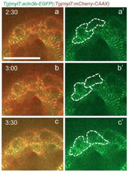
Sarcomere reassembly after cardiomyocyte cytokinesis. Follow up of the time-lapse live imaging of the beating Tg(myl7:actn3b-EGFP); Tg(myl7:mCherry-CAAX) heart shown in Figure 3H. The two daughter cells reassemble their sarcomeres (a-b'), and seem to recover the striated sarcomeric pattern approximately 3 hours after the disassembly was first observed in the dividing cardiomyocyte (c-c'). Scale bar, 20 µm. |
Expression Data
Expression Detail
Antibody Labeling
Phenotype Data
Phenotype Detail
Acknowledgments
This image is the copyrighted work of the attributed author or publisher, and
ZFIN has permission only to display this image to its users.
Additional permissions should be obtained from the applicable author or publisher of the image.
Full text @ Development