
FIGURE
Fig. S1
Fig. S1
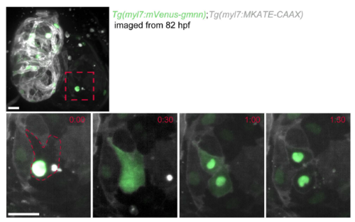
myl7:mVenus-Gmnn signal diffuses throughout the cardiomyocyte prior to cytokinesis. Zoomed image of an atrial cardiomyocyte in a 82 hpf Tg(myl7:mVenus-gmnn); Tg(myl7:MKATE-CAAX) heart acquired using a lightsheet microscope time-lapse for every 30 minutes. Diffusion of the fluorescent signal throughout the cardiomyocyte is observed before it divides. Once cytokinesis is completed, the myl7:mVensu-Gmnn signal is again restricted to the nuclei and then disappears within 1 hour. Scale bars, 20 µm. |
Expression Data
Expression Detail
Antibody Labeling
Phenotype Data
Phenotype Detail
Acknowledgments
This image is the copyrighted work of the attributed author or publisher, and
ZFIN has permission only to display this image to its users.
Additional permissions should be obtained from the applicable author or publisher of the image.
Full text @ Development