
FIGURE
Fig. S2
Fig. S2
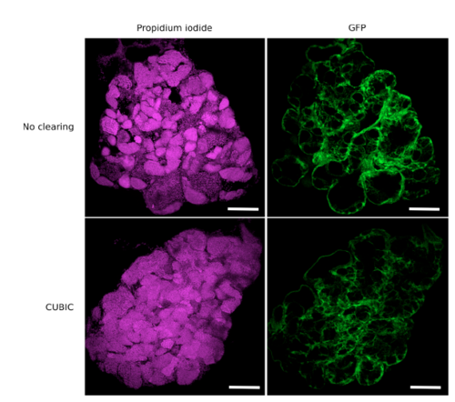
Confocal imaging of transverse vibratome sections of CUBIC-‐ cleared and uncleared testes. Testes were dissected from the zebrafish transgenic line Tg(gsdf:GFP), cleared or not with CUBIC and stained with propidium iodide. Transverse sections were performed with a vibratome to assess the nuclear staining in depth. Confocal images show that all nuclei in the center of the sample are efficiently stained. Images correspond to maximal projections of 10 optical slices (z-‐step = 3 μm). Nuclei are in magenta and Sertoli GFP cells in green. Scale bar: 100 μm. |
Expression Data
Expression Detail
Antibody Labeling
Phenotype Data
Phenotype Detail
Acknowledgments
This image is the copyrighted work of the attributed author or publisher, and
ZFIN has permission only to display this image to its users.
Additional permissions should be obtained from the applicable author or publisher of the image.
Full text @ Sci. Rep.