
Fig. 3
- ID
- ZDB-FIG-180827-15
- Publication
- Ferrero et al., 2018 - Embryonic Microglia Derive from Primitive Macrophages and Are Replaced by cmyb-Dependent Definitive Microglia in Zebrafish
- Other Figures
- All Figure Page
- Back to All Figure Page
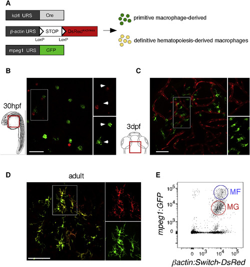
Embryonic and Adult MG Have Distinct Origins (A) Scheme of the transgenic lines used to address the MG potential of primitive versus definitive hematopoiesis. (B and C) Whole-mount fluorescence microscopy images of Tg(kdrl:Cre; ßactin:Switch-DsRed; mpeg1:GFP) triple-transgenic embryos show absence of DsRed expression (red) in GFP+ pMFs (green) on the yolk ball at 30 hpf (B), and in GFP+ embryonic MG (green) in the brain parenchyma at 3 dpf (C) (n = 14). (D) In contrast, in sections of adult triple-transgenic animals, DsRed labeling is observed in all GFP+ MG cells. (E) Flow cytometry analysis on a brain dissected from a 12-week-old triple-transgenic animal identifies blood-derived mpeg1:GFPhigh CNS-associated MFs (blue circle, MFs) and mpeg1:GFPlow MG (red circle, MG). Both populations express the DsRed reporter at high level (n = 5). Scale bar: 50 μm. MFs, macrophages; MG, microglia. See also Figures S4 and S5. |
| Genes: | |
|---|---|
| Fish: | |
| Anatomical Terms: | |
| Stage Range: | Prim-15 to Adult |