
Fig. 1
- ID
- ZDB-FIG-180827-13
- Publication
- Ferrero et al., 2018 - Embryonic Microglia Derive from Primitive Macrophages and Are Replaced by cmyb-Dependent Definitive Microglia in Zebrafish
- Other Figures
- All Figure Page
- Back to All Figure Page
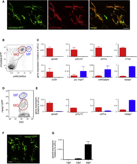
MG Identity Is Conserved in the Zebrafish (A) Fluorescence of GFP (left panel) and DsRed (middle panel) in the brain of adult cd45:DsRed; mhc2dab:GFP double-transgenic fish. The right panel shows a merge of both fluorescent channels (n = 3). Scale bar: 10 μm. (B) Flow cytometry analysis on brain cell suspensions from adult Tg(cd45:DsRed; mhc2dab:GFP) identifying cd45:DsRedlowmhc2dab:GFP+ MG (red gate, MG) and cd45:DsRedhighmhc2dab:GFP+ CNS-associated macrophages (blue gate, MFs). (C) Comparison of the relative expression of apoeb, p2ry12, csf1ra, C1qc, csf3r, mpeg1, pu.1/spi1b, and cd45/ptprc transcripts between MG (red bars) and MFs (blue bars) cell subsets isolated by FACS. Error bars represent mean ± SD (n = 3). (D and E) Flow cytometry of a brain cell suspension from adult mpeg1:GFP transgenics (D) and expression of apoeb, p2ry12, csf1ra, and mpeg1 by qPCR in FACS-sorted mpeg1:GFP+ cell subsets (E). Error bars represent mean ± SD (n = 3). (F) GFP immunohistochemistry on adult mpeg1:GFP brain sections. Scale bar: 25 μm. (G) apoeb expression in mpeg1:GFP+ cells sorted at 1, 2, and 3 dpf. MG, microglia; MFs, macrophages. |
| Genes: | |
|---|---|
| Fish: | |
| Anatomical Terms: | |
| Stage: | Adult |