
Fig. 7
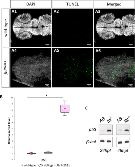
Massive p53-dependent apoptosis in the fbl mutant. (A) Horizontal optic sections of TUNEL labeling at 24 hpf in wild-type (A1-A3) and mutant (A4-A6) embryos. Gray: DAPI staining, Green: TUNEL staining. Scale bar: 50 µm. Anterior is to the left. (B) RT-qPCR quantification of relative levels of tp53 mRNA at 72 hpf shows a strong increase in tp53 expression in mutants. Purple: wild-type, gray: siblings, pink: mutants. Statistical analyses were performed on biological triplicates, p-value (Kruskal-Wallis test): 0.049. (C) RT-PCR for tp53 in 24 hpf and 48 hpf mutant embryos showing a large increase in tp53 expression. Anterior is to the left. |
| Gene: | |
|---|---|
| Fish: | |
| Anatomical Term: | |
| Stage Range: | Prim-5 to Protruding-mouth |
| Fish: | |
|---|---|
| Observed In: | |
| Stage Range: | Prim-5 to Protruding-mouth |
Reprinted from Developmental Biology, 437(1), Bouffard, S., Dambroise, E., Brombin, A., Lempereur, S., Hatin, I., Simion, M., Corre, R., Bourrat, F., Joly, J.S., Jamen, F., Fibrillarin is essential for S-phase progression and neuronal differentiation in zebrafish dorsal midbrain and retina, 1-16, Copyright (2018) with permission from Elsevier. Full text @ Dev. Biol.