
|
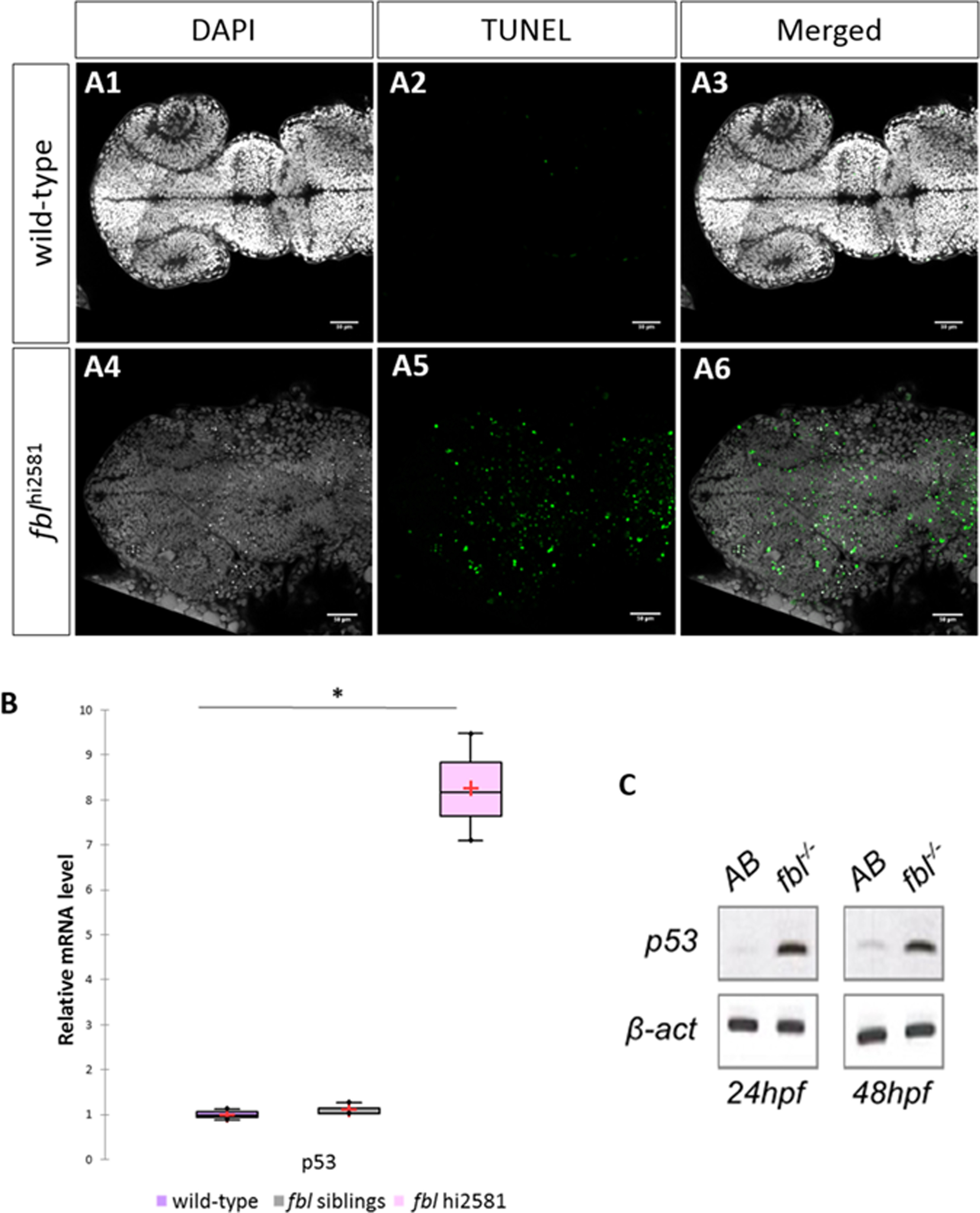
Fig. 7
Massive p53-dependent apoptosis in the fbl mutant. (A) Horizontal optic sections of TUNEL labeling at 24 hpf in wild-type (A1-A3) and mutant (A4-A6) embryos. Gray: DAPI staining, Green: TUNEL staining. Scale bar: 50 µm. Anterior is to the left. (B) RT-qPCR quantification of relative levels of tp53 mRNA at 72 hpf shows a strong increase in tp53 expression in mutants. Purple: wild-type, gray: siblings, pink: mutants. Statistical analyses were performed on biological triplicates, p-value (Kruskal-Wallis test): 0.049. (C) RT-PCR for tp53 in 24 hpf and 48 hpf mutant embryos showing a large increase in tp53 expression. Anterior is to the left.
Reprinted from Developmental Biology, 437(1), Bouffard, S., Dambroise, E., Brombin, A., Lempereur, S., Hatin, I., Simion, M., Corre, R., Bourrat, F., Joly, J.S., Jamen, F., Fibrillarin is essential for S-phase progression and neuronal differentiation in zebrafish dorsal midbrain and retina, 1-16, Copyright (2018) with permission from Elsevier. Full text @ Dev. Biol.