
FIGURE
Fig. 5-S1
Fig. 5-S1
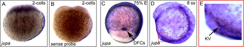
jupa is maternally supplied and enriched in precursor dorsal forerunner cells (DFCs) and KV. (A–E) RNA in situ hybridizations of jupa during early zebrafish development. (A) Antisense jupa probe revealed maternal jupa mRNA in 2 cell stage embryo. (B) A control jupa sense probe showed no staining. (C–D) jupa mRNA was detected in DFCs (black arrow) and KV cells (red square) during epiboly and early somite stages. (E) Enlarged region depicting jupa staining in KV (black arrow). |
Expression Data
Expression Detail
Antibody Labeling
Phenotype Data
Phenotype Detail
Acknowledgments
This image is the copyrighted work of the attributed author or publisher, and
ZFIN has permission only to display this image to its users.
Additional permissions should be obtained from the applicable author or publisher of the image.
Full text @ Elife