FIGURE

Fig. S4

ID
ZDB-FIG-180226-20
Publication
Cao et al., 2017 - Tension Creates an Endoreplication Wavefront that Leads Regeneration of Epicardial Tissue
Other Figures
All Figure Page
Back to All Figure Page
Fig. S4

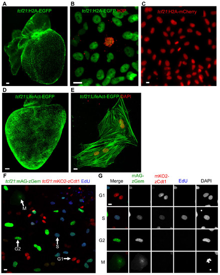
New Reporter Lines for Live Imaging (Related to Figure 3)
(A, B) A tcf21:H2A-EGFP reporter line indicates chromatin in epicardial cells of a wholemounted heart (A) and in epicardial explant culture (B). Anti-phospho-Histone H3 staining (H3P) was performed to label condensed chromatin (red). Scale bars, 100 μm (A) or 10 μm (B).
(C) High-magnification view of tcf21:H2A-mCherry epicardial cells. Scale bar, 10 μm
(D, E) A tcf21:LifeAct-EGFP reporter labels F-actin in whole-mounted (D) or isolated epicardial cells (E). DAPI stains nuclei red in (E). Scale bars, 100 μm (D) or 10 μm (E).
(F, G) Epicardial explant culture from a tcf21:FUCCI heart, stained after a 1 h pulse of EdU to mark S phase. Arrows in (F) denotes different cell cycle phases, also highlighted in (G). Scale bars, 10 μm.

Expression Data

Expression Detail
Antibody Labeling
Phenotype Data

Phenotype Detail
Acknowledgments
This image is the copyrighted work of the attributed author or publisher, and ZFIN has permission only to display this image to its users. Additional permissions should be obtained from the applicable author or publisher of the image.

Reprinted from Developmental Cell, 42, Cao, J., Wang, J., Jackman, C.P., Cox, A.H., Trembley, M.A., Balowski, J.J., Cox, B.D., De Simone, A., Dickson, A.L., Di Talia, S., Small, E.M., Kiehart, D.P., Bursac, N., Poss, K.D., Tension Creates an Endoreplication Wavefront that Leads Regeneration of Epicardial Tissue, 600-615.e4, Copyright (2017) with permission from Elsevier. Full text @ Dev. Cell