FIGURE

Fig. 4

ID
ZDB-FIG-180226-12
Publication
Cao et al., 2017 - Tension Creates an Endoreplication Wavefront that Leads Regeneration of Epicardial Tissue
Other Figures
All Figure Page
Back to All Figure Page
Fig. 4

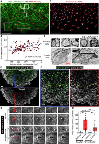
Leader Cells Display Higher Migration Velocity and Mechanical Tension than Followers


(A) Epicardial explant culture showing F-actin and nuclei by tcf21:LifeAct-EGFP;tcf21:H2A-mCherry reporters. The framed regions were enlarged for display in (D). LifeAct-EGFP is shown in green and H2A-mCherry in red. Scale bars, 100 μm.


(B) Same experiment as in (A) showing velocity vectors (white arrows) for each nucleus (H2A-mCherry, red) in a 5-hr window. Scale bars, 100 μm.


(C) Dot plot for each individual nucleus indicating the average speed in 15.5 hr over the average distance from the leading edge. The blue line indicates the regression result. n = 224.


(D) Magnified view of the framed regions in (A). LifeAct-EGFP is shown in inverted grayscale. Scale bars, 20 μm.


(E and F) A 5-day tcf21:H2A-EGFP (green) epicardial explant culture was stained with pMLC (Ser19, grayscale). The framed regions in (E) are enlarged to show details in (F). The yellow dashed lines approximately separate follower (top) and leader cell regions (bottom). Scale bars, 100 μm.


(G) Video frames of explanted epicardial cells from a tcf21:LifeAct-EGFP line showing laser incisions and recoil of the cells. LifeAct-EGFP is shown in grayscale. Red double arrows indicate position and length of incisions. F, follower cells; L, leader cells. Scale bar, 20 μm.


(H) Quantification of the initial recoil velocity of leader and follower cells after cutting in a direction parallel or perpendicular to the migration direction. n = 12 (parallel-follower), 16 (parallel-leader), 14 (perpendicular-follower), and 15 (perpendicular-leader), respectively. ∗∗∗p < 0.001; ns, not significant; Mann-Whitney rank-sum test. Error bars indicate SD.


See also Movies S4 and S6.

Expression Data

Expression Detail
Antibody Labeling
Phenotype Data

Phenotype Detail
Acknowledgments
This image is the copyrighted work of the attributed author or publisher, and ZFIN has permission only to display this image to its users. Additional permissions should be obtained from the applicable author or publisher of the image.

Reprinted from Developmental Cell, 42, Cao, J., Wang, J., Jackman, C.P., Cox, A.H., Trembley, M.A., Balowski, J.J., Cox, B.D., De Simone, A., Dickson, A.L., Di Talia, S., Small, E.M., Kiehart, D.P., Bursac, N., Poss, K.D., Tension Creates an Endoreplication Wavefront that Leads Regeneration of Epicardial Tissue, 600-615.e4, Copyright (2017) with permission from Elsevier. Full text @ Dev. Cell