
Fig. 4
- ID
- ZDB-FIG-180131-15
- Publication
- Escobar-Aguirre et al., 2017 - Microtubule-actin crosslinking factor 1 (Macf1) domain function in Balbiani body dissociation and nuclear positioning
- Other Figures
- All Figure Page
- Back to All Figure Page
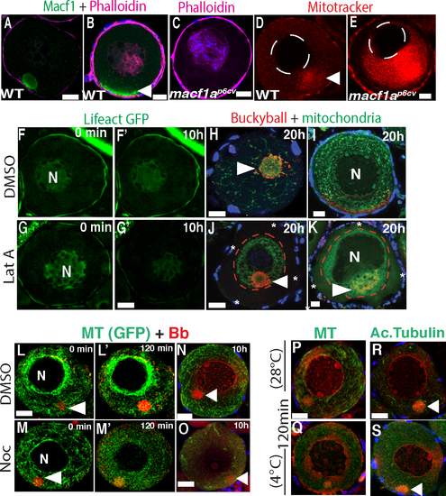
Effect of disrupting actin and MTs on the Bb and nuclear positioning. A-B) Macf1 (green) and phalloidin staining in WT, and C) phalloidin in macf1ap6cv mutant oocytes labeling cortical and intranuclear actin. N = 3 ovaries, 5 WT and 10 macf1ap6cv mutant oocytes. WT (D’) and macf1ap6cv mutant (E) live stage I oocytes stained with MitoTracker to visualize the cortical detachment of mitochondria in macf1a mutant. N = 3 ovaries, 22 WT and 7 macf1ap6cv oocytes. F-G) Live imaging of Lifeact-GFP ovaries treated with DMSO (F-F’) or LatA (G-G’) for 10h. G’) GFP signal decreases indicating acting disruption. H-K) Dissected ovaries enriched for stage 1 and 2 oocytes were treated with DMSO (H-I) or LatA (J-K) for 20 h, then fixed and stained for mitochondria (DiOC6, green) and Buc (red). Asterisks and red dotted line mark the cortical detachment of mitochondria and Buc in LatA treated oocytes (J-K). The number of oocytes imaged that showed the mitochondrial cortical detachment in DMSO and LatA treated was 3/25 and 25/40, respectively. F) The oocytes that displayed an acentric nucleus after LatA treatment was 15 of 40 oocytes. N ≥ 5 ovaries. L-M) live imaging of stage I oocytes Tg(ef1a:dclk-GFP) to visualize MTs (green) and Mitotracker (mitochondria, red) to visualize the Bb. Oocytes were treated with DMSO (L, control) or nocodazole (M). N≥5 ovaries, 75 DMSO and 73 nocodazole treated oocytes. N-O) Ovaries from Tg (EMTB-3GFP) treated with DMSO (N) or nocodazole (O) for 10h, and stained for MTs (GFP) and mitochondria (DiOC₆). N = 3 ovaries, 10 oocytes DMSO treated and 11 nocodazole treated. P-S) Ovaries incubated at 28°C (P,R) or 4°C (Q,S) to depolymerize stable MTs, were fixed after 120 min cold treatment and stained for MTs (P, Q; Tg(ef1a:dclk-GFP)) or acetylated tubulin (R, S). N≥5 ovaries, 53 (28°C) and 50 (4°C) oocytes. Arrowheads indicate the Bb. Scale bar: 20 μm. |
| Genes: | |
|---|---|
| Antibody: | |
| Fish: | |
| Conditions: | |
| Anatomical Terms: | |
| Stage: | Adult |
| Fish: | |
|---|---|
| Conditions: | |
| Observed In: | |
| Stage: | Adult |