
Fig. 3
- ID
- ZDB-FIG-180131-14
- Publication
- Escobar-Aguirre et al., 2017 - Microtubule-actin crosslinking factor 1 (Macf1) domain function in Balbiani body dissociation and nuclear positioning
- Other Figures
- All Figure Page
- Back to All Figure Page
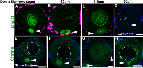
Localization of Macf1a in stage I oocytes. A-C) Macf1a immunostaining (green) in stage I oocytes shows localization nuclearly and within the Bb (A), and later at the cortex during Bb disassembly (B, C). D) Macf1a staining was negative in a macf1asa12708 mutant oocyte. E-G) GFP staining in Gt(macf1a–citrine)ct68a line also showed Macf1a-Citrine localization to the Bb and at the cortex during disassembly. DAPI (blue/magenta) stains DNA. A-D) N≥ 3 ovaries, 20 isolated WT oocytes were examined (see methods), 16 oocytes showed Macf1 nuclear staining; 18 macf1asa12708 oocytes were examined. E-H) N = 3 ovaries, 9 of 20 Gt(macf1a–citrine)ct68a/+ oocytes showed nuclear staining (H). Representative images from 3 experiments. Dotted white lines outline the nucleus. Images are a sum of 3 single optical confocal sections. Arrowheads indicate the Bb. Scale bars: 20 μm. |
| Genes: | |
|---|---|
| Fish: | |
| Anatomical Terms: | |
| Stage: | Adult |
| Fish: | |
|---|---|
| Observed In: | |
| Stage: | Adult |