
Fig. 2
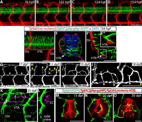
VTAs serve as critical scaffolds for subsequent vascularization around and within the spinal cord. (A–D) TgBAC(gfap:gfap-EGFP);Tg(kdrl:ras-mcherry) trunks at 76 (A), 102 (B), 124 (C), and 154 (D) hpf show a developmental time course of VTA formation. (E and F–F″) A 154 hpf TgBAC(gfap:gfap-EGFP);Tg(kdrl:ras-mcherry) trunk (E) and its sections counterstained with DAPI (F–F″). Radial glia endfeet and VTAs are physically adjacent (white arrows). High-magnification image of the region inside the dashed line (F′) is shown in F″. (G–I) Tg(kdrl:EGFP) trunk vasculature at different developmental stages. At 7 dpf, most VTAs are formed (G, yellow arrows). At 11 dpf, sprouting vessels emerge between dorsal ISVs (H, yellow arrows), and these vessels have established connections with neighboring VTAs or ISVs by 15 dpf (I). (J) High-magnification image of Tg(kdrl:EGFP) trunk vasculature at 18 dpf. Vessels around the spinal cord are connected to VTAs (yellow arrows). (K and L) z-stack projection images of 25 dpf Tg(kdrl:EGFP) trunk vasculature. Blood vessels inside the spinal cord are shown in magenta. Pink arrows point to the sites where angiogenic sprouting into the spinal cord takes place. It mostly occurs from the dorsolateral sides of the PNVP. (M–O) Tg(gfap:NTR);Tg(kdrl:EGFP) trunk sections at different developmental stages. At 15 dpf, blood vessels are not observed inside the spinal cord (M). Blood vessel invasion into the spinal cord is observed by 20 dpf (N). Increased number and density of spinal cord vessels are observed at 30 dpf (O). (Scale bars, 100 µm in D and G–K, 50 µm in E and M–O, and 20 µm in F and F′.) |
| Genes: | |
|---|---|
| Fish: | |
| Anatomical Terms: | |
| Stage Range: | Protruding-mouth to Days 30-44 |