
Fig. S5
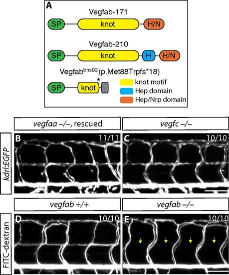
VTA formation in vegfaabns1, vegfabbns92, and vegfchu6410 mutants. (A) Predicted domain structure of WT Vegfab isoforms -171, -210, and Vegfabbns92. Vegfab-171 and -210 consist of a signal peptide (SP), a knot motif, and heparin (Hep) and/or Hep/Nrp1 (Nrp) binding domains. vegfabbns92 encodes a truncated peptide (p.Met88Trpfs*18). (B and C) 8 dpf Tg(hsp70l:vegfaa165) vegfaa−/− (B) and vegfc−/− (C) larval trunk vasculature visualized by Tg(kdrl:EGFP) expression. Tg(hsp70l:vegfaa165) vegfaa−/− embryos were heat-shocked at 10 and 24 hpf to rescue their early vascular phenotype. VTAs are present in all of the vegfaa−/− (B; n = 11) and vegfc−/− (C; n = 10) larvae examined. (D and E) 8 dpf vegfab+/+ (D) and vegfab−/− (E) larval trunk vasculature visualized by FITC-dextran microangiography. vegfab−/− larvae lack lumenized VTAs (yellow arrows, E). |