
Fig. S8
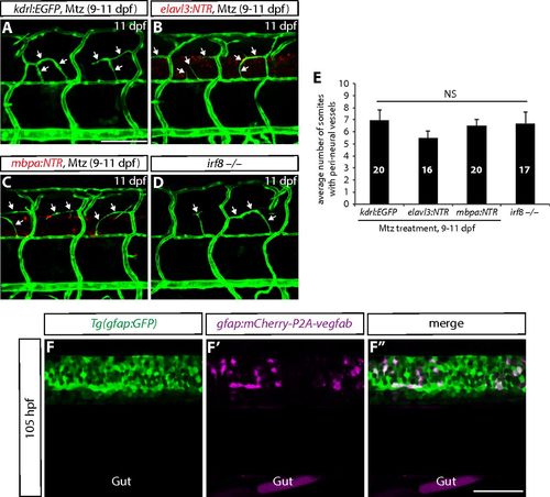
Ablation of neurons, oligodendrocytes, or microglia does not lead to compromised PNVP formation; specific expression of gfap:mCherry-P2A-Vegfab in Tg(gfap:GFP)+ radial glia. (A–D) 11 dpf Tg(kdrl:EGFP) (A), Tg(elavl3:NTR);Tg(kdrl:EGFP) (B), and Tg(mbpa:NTR);Tg(kdrl:EGFP) (C) larval trunk vasculature after treatment with Mtz from 9 to 11 dpf. 11 dpf Tg(kdrl:EGFP) irf8−/− trunk vasculature is shown in D. Mtz-treated control fish (A) as well as larvae in which neurons (B), oligodendrocytes (C), or microglia (D) were depleted exhibit vessels (white arrows) that emerge between the dorsal ISVs around the spinal cord. (E) Quantification of the average number of somites with peri-neural blood vessels (10 somites examined per animal). Quantification was performed at 11 dpf. Values represent means ± SEM. (F–F″) 105 hpf trunk of Tg(gfap:GFP) fish injected with the gfap:mCherry-P2A-vegfab plasmid. Injection of the gfap:mCherry-P2A-vegfab plasmid drives mCherry expression in cells within the spinal cord (F′), which are also positive for Tg(gfap:GFP) expression (F). All mCherry+ cells are also GFP+ (F″). (Scale bars, 100 µm in A and 50 µm in F″.) |