
FIGURE
Fig. S6
- ID
- ZDB-FIG-171108-53
- Publication
- Fei et al., 2016 - Cardiac Light-Sheet Fluorescent Microscopy for Multi-Scale and Rapid Imaging of Architecture and Function
- Other Figures
- All Figure Page
- Back to All Figure Page
Fig. S6
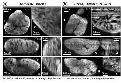
Imaging comparison between c-LSFM and confocal microscopy using 120 dpf zebrafish hearts. (a) The original x-y plane image, reconstructed x-z and y-z plane images obtained from confocal data. (b) The original x-y plane image, reconstructed x-z and y-z plane images obtained from c-LSFM data. The zoomed-in images shown in the right columns (2, 4, 6) indicate the lateral and axial resolving powers of confocal and c-LSFM. |
Expression Data
Expression Detail
Antibody Labeling
Phenotype Data
Phenotype Detail
Acknowledgments
This image is the copyrighted work of the attributed author or publisher, and
ZFIN has permission only to display this image to its users.
Additional permissions should be obtained from the applicable author or publisher of the image.
Full text @ Sci. Rep.