
Fig. S3
- ID
- ZDB-FIG-171108-51
- Publication
- Fei et al., 2016 - Cardiac Light-Sheet Fluorescent Microscopy for Multi-Scale and Rapid Imaging of Architecture and Function
- Other Figures
- All Figure Page
- Back to All Figure Page
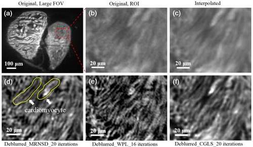
Post-image processing of adult zebrafish heart with resolution enhancement by different deconvolution techniques. (a) Zebrafish atrium and ventricle are visualized on one section of raw image. (b) The individual cardiomyocytes were unresolvable from the selected region-of-interest from the atrium due to the optics blurring and under-sampling by the camera. (c) The image was first scaled up with 3X b-spline interpolation to partially recover the information loss from incomplete sampling. (d), (e) and (f) illustrate the interpolated images deblurred by iterative MRNSD, WPL and CGLS deconvolution, respectively. The WPL algorithm generates most effective deblurring to recover sharp and high frequency signals. However, it also generates image discontinuity, likely due to the application of wiener filter. The CGLS algorithm appears to be mild, generating the least degree of deblurring. Of the three resolution enhancement algorithms, the MRNSD method provides optimal trade-off between the resolution enhancement and information preservation. |