
Fig. 4
- ID
- ZDB-FIG-171108-43
- Publication
- Fei et al., 2016 - Cardiac Light-Sheet Fluorescent Microscopy for Multi-Scale and Rapid Imaging of Architecture and Function
- Other Figures
- All Figure Page
- Back to All Figure Page
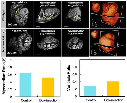
Rapid 3-D images to recapitulate trabeculated network in response to doxorubicin (Dox) treatment in the adult zebrafish. (a) A representative wild-type zebrafish heart at 120 dpf. The coronal, sagittal and transverse planes of the heart displayed a compact trabecular network. The atrioventricular valve (AV) was identified (yellow arrows). Scale bars are 200 μm in length. In the rightmost column, a 3-D rendering of the “digital heart” was reconstructed by stacking 500 slices of plane images in volume. The 3-D structure of the “digital heart” can be assessed by arbitrary cropping. (b) A representative Dox-injected zebrafish heart at 120 dpf. The endocardial cavity appeared enlarged and the trabecular network was accentuated. (c) The quantified volume ratios of the myocardium (left) and the ventricle cavity (right) in the whole heart. |