
FIGURE
Fig. S5
- ID
- ZDB-FIG-171108-52
- Publication
- Fei et al., 2016 - Cardiac Light-Sheet Fluorescent Microscopy for Multi-Scale and Rapid Imaging of Architecture and Function
- Other Figures
- All Figure Page
- Back to All Figure Page
Fig. S5
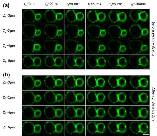
Comparison between prior to and post 4-D synchronization algorithm. (a) Before synchronization, zebrafish cardiac contractions at different Z positions were not in the same stage. (b) After synchronization, all Z positions were synchronized in the same cardiac contraction stage. Therefore, stacked images for 3-D reconstruction at certain time points were obtained and provided volume information. Scale bar = 50µm. |
Expression Data
Expression Detail
Antibody Labeling
Phenotype Data
Phenotype Detail
Acknowledgments
This image is the copyrighted work of the attributed author or publisher, and
ZFIN has permission only to display this image to its users.
Additional permissions should be obtained from the applicable author or publisher of the image.
Full text @ Sci. Rep.