
Fig. S5
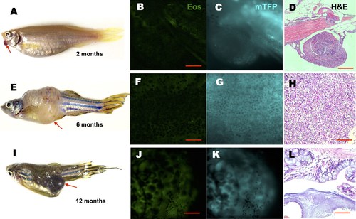
Representative tumors induced by constitutive kRASG12V activation. Tg(ubi:Cre- ERT2) fish injected with ubi:loxP-Eos-stop-loxP-kRASG12V-T2A-mTFP plasmid were incubated at 1dpf in cyclofen or caged cyclofen + UV. Various tumors (noted by arrows) were observed in fish at 2 (A), 6 (E) and 12 (I) months. The tumors expressed reduced EosFP (B, F, J), and were characterized by both strong expression of mTFP (C, G, K) and histopathological analysis (D, H, L). Condensed nuclei and distorted cell shapes are typical of tumor morphologies, however the different overall textures in D, H and L suggest different tumor types. Scale bar: B, D, F, G, J, 400 μm; D, H 200 μm; L 50 μm. |