
Fig. S6
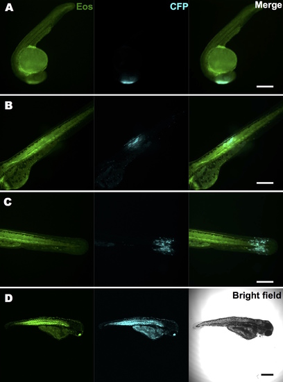
Local transient activation of oncogene expression with a UV laser at different stages and in various tissues. We crossed a stable Tg(ubi:Eos;UAS:kRASG12V-T2A-CFP) line with a Tg(ubi:Gal4-ERT2) line. Embryos were pre-incubated in 4 μM caged cyclofen for 4 hours. They were illuminated at 10 hpf (A) or 32 hpf (B, C) with the 405 nm line of a LEICA SP5-BLUE microscope for 5 seconds. Fluorescent images were taken after 18 hours and showed precise spatial control of oncogene activation. While all locally illuminated embryos developed normally, many globally illuminated fish (D) exhibited severe developmental defects and died. Scale bar: 400 μm. |