
Fig. 5
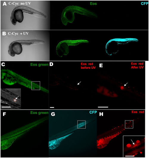
KRASG12V activation and tracking in a small group of cells within a live zebrafish embryo. A stable Tg(ubi:Eos;UAS:kRASG12V-T2A-CFP) line was generated and crossed with the Tg(ubi:Gal4-ERT2) line. Caged cyclofen alone at 1 dpf was not able to induce CFP expression (A), while 2-minute UV illumination (B) effectively turned on CFP after 18 hours. (C) Local activation of kRASG12V expression was achieved by introducing a focalized UV beam onto embryos pre-incubated with caged cyclofen. (D,E) 1 minute UV effectively converted EosFP from green to red. (F,G) After 18 hours, CFP expressed in a localized region of the illuminated embryo and weak remaining EosFP red signal was detected in the same region (H). Scale bar: 200 μm. |