
Fig. 1
- ID
- ZDB-FIG-170922-25
- Publication
- Walsh et al., 2017 - Progranulin regulates neurogenesis in the developing vertebrate retina
- Other Figures
- All Figure Page
- Back to All Figure Page
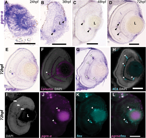
Expression of pgrn-a in the developing retina. (A–D) Cross-sections of the retina following whole mount in situ hybridizations (ISHs) showing pgrn-a expression in WT at 24 (A), 36 (B), 48 (C), and 72 hpf (D). (E–L) pgrn-a is microglia-specific by 72 hpf. pgrn-a ISH (E + G) followed by immunohistochemistry of L-plastin (F) and 4C4 (H) showing colocalization of pgrn-a and microglial markers (arrowheads). Double ISH in the same section showing DAPI (I), pgrn-a (J), and fms (K) expression, and overlay (L) showing colocalization of pgrn-a and fms in the retina (arrowheads). In panel D, it appears there is transcript expression in the CMZ. However, there is no evidence for this following whole mount ISH (see Supporting Information Fig. S1), and we infer that in sections this apparent labeling is spurious. Outer nuclear layer (ONL); inner nuclear layer (INL); and ganglion cell layer (GCL); (L) lens. Scale bar equals 50 µm. |