
Fig. 5
- ID
- ZDB-FIG-170815-18
- Publication
- Lim et al., 2017 - Caveolae Protect Notochord Cells against Catastrophic Mechanical Failure during Development
- Other Figures
- All Figure Page
- Back to All Figure Page
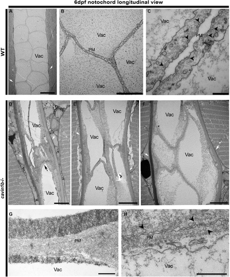
Longitudinal Ultrastructure of 6-dpf cavin1b−/− Notochords (A–C) Electron micrographs of a notochord longitudinal section from a 6-dpf WT embryo. Magnified view in (B) shows tightly apposed neighboring notochordal cells. (C) depicts neighboring notochord cells and caveolae. (D–F) Longitudinal electron micrographs of notochord regions in lesion-rich sites from 6-dpf cavin1b−/− embryos, each image representing a different zebrafish. Note aberrantly shaped vacuoles and disrupted cell morphology. For correlative light microscopy image of lesion presented in (D), refer to Figure S4F. (G and H) Longitudinal electron micrographs of cavin1b−/− cell-cell septum between notochord cells showing a prominent reduction in caveola number. Black arrow, notochord lesion; black arrowhead, caveola; white arrow, perinotochordal sheath. The scale bars represent (A, B, and D–F) 10 μm and (C, G, and H) 500 nm. |
| Fish: | |
|---|---|
| Observed In: | |
| Stage: | Day 6 |