
Fig. 1
- ID
- ZDB-FIG-170815-14
- Publication
- Lim et al., 2017 - Caveolae Protect Notochord Cells against Catastrophic Mechanical Failure during Development
- Other Figures
- All Figure Page
- Back to All Figure Page
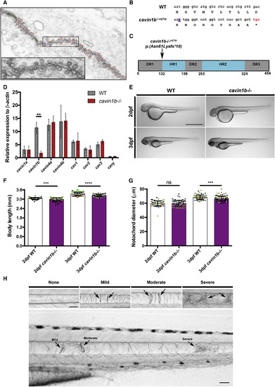
Mutant cavin1b−/− Zebrafish Exhibit Notochord Lesions (A) Electron micrograph depicting dense caveolae in the septum between two neighboring zebrafish embryonic notochord cells. Superimposed schematic trace highlights abundant caveolae (red), plasma membrane (blue), and junctions (green). The inset shows magnification of highlighted area. (B) Alignment of nucleotide and deduced amino acid sequences from WT and cavin1b−/− lines. An adenine insertion in the mutant sequence is highlighted in purple and underlined. Asterisk (∗) indicates the truncating stop codon in the mutant sequence (marked in red). See Figures S1A–S1E for cavin1b CRISPR/Cas9 mutant generation and genotyping. (C) Cavin1b protein domains (schematic). Arrow indicates the site of predicted truncation of selected mutant. The DR (disordered region) and HR (helical region) protein regions are boxed in gray and blue, respectively. Standard sequence mutation nomenclature is used [6]. (D) mRNA expression levels of caveola-associated genes by qRT-PCR (relative to β-actin). 5-dpf WT and cavin1b−/− embryos are shown (n = 3 clutches; performed in triplicate). See Figure S1F for related RT-PCR gel. ∗∗p ≤ 0.01; two-way ANOVA with Tukey’s multiple-comparison test. Data are presented as mean ± SD. See Table S1 for primer details. (E) Gross morphology of live 2- and 3-dpf WT and cavin1b−/− embryos (treated with 1-phenyl-2-thiourea [PTU] to inhibit melanization). The scale bar represents 1 mm. (F) Body length (mm) in 2- and 3-dpf WT and cavin1b−/− embryos (2 dpf: n = 62 [WT] and n = 93 [mutant]; 3 dpf: n = 76 [WT] and n = 100 [mutant]). Four clutches per group are shown. Colored dots indicate different clutches. (G) Notochord diameter (μm) of 2- and 3-dpf WT and cavin1b−/− embryos (2 dpf: n = 65 [WT] and n = 94 [mutant]; 3 dpf: n = 79 [WT] and n = 100 [mutant]). Four clutches per group are shown. For observations on live chordamesoderm transition, see Figure S2A. For characterization of notochord vacuole formation, see Figures S2B–S2F. For (F)–(G), ns, p > 0.05; ∗∗∗p ≤ 0.001; ∗∗∗∗p ≤ 0.0001; two-tailed t tests. Data are presented as mean ± SD. (H) Notochord lesions. (Top row) Notochord lesions of varying severity (mild, moderate, or severe) are shown. (Bottom row) Representative image of a 3-dpf cavin1b−/− notochord possessing all three qualities of lesion severity is shown. The scale bar represents 50 μm. See Figures S1G and S1H for lesions in another cavin1b mutant line. |
| Gene: | |
|---|---|
| Fish: | |
| Anatomical Term: | |
| Stage: | Day 5 |
| Fish: | |
|---|---|
| Observed In: | |
| Stage Range: | Long-pec to Day 5 |