
Fig. S1
- ID
- ZDB-FIG-170306-9
- Publication
- Gays et al., 2017 - An exclusive cellular and molecular network governs intestinal smooth muscle cell differentiation in vertebrates
- Other Figures
- All Figure Page
- Back to All Figure Page
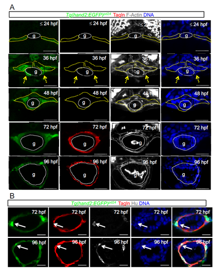
Time course analyses of Tg(hand2:EGFP)pd24 and iSMC marker expression during intestine development. a) Time course analyses of Tg(hand2:EGFP)pd24 and iSMC marker expression (Tagln) during intestine development. Confocal transverse sections of the gut region between the somites 7 and 13 of Tg(hand2:EGFP)pd24 embryos from 24 hpf to 96 hpf as indicated and stained with phalloidin (grey) and Tagln (red). Enteric endoderm is highlight with dashed white line. The yellow dashed line highlights LPM/hand2+ cells. These panels are referring to Figure 1a. Blue = nuclei. Scale bar, 30 μm. Gut: g; LPM: lateral plate mesoderm. b) LPM-derived enteric neurons show high levels of hand2 expression. Confocal transverse sections of the gut region between the somites 7 and 13 of Tg(hand2:EGFP)pd24 embryos stained for the enteric neuron marker Hu (gray) and Tagln (red) at 72 and 96 hpf. Select LPM/hand2+ cells express strong levels of hand2 and become enteric neurons (arrows). Blue = nuclei. Scale bar, 20 μm. |