
Fig. 1
- ID
- ZDB-FIG-160729-4
- Publication
- Lv et al., 2016 - Synaptic Ribbons Require Ribeye for Electron Density, Proper Synaptic Localization, and Recruitment of Calcium Channels
- Other Figures
- All Figure Page
- Back to All Figure Page
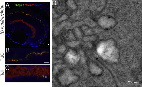
Ribeye and Synaptic Ribbons Remain in the Retina of ribeye a(Δ10)/ ribeye b(Δ7) (re[a(Δ10)/b(Δ7)]) double homozygous mutants. (A) Confocal image of Ribeye b (green) and post-synaptic maker MAGUK (red) staining in 5-dpf re[a(Δ10)/b(Δ7)] homozygous mutants. DAPI (blue) stains for the nucleus. Scale bar, 5 µm. (B) 3× magnification of outer plexiform layer staining in 5-dpf re[a(Δ10)/b(Δ7)] homozygous mutants retina. Scale bar, 5 µm. (C) 3× magnification of inner plexiform layer staining in 5-dpf re[a(Δ10)/b(Δ7)] homozygous mutants retina. Scale bar, 5 µm. (D) Electron micrograph of photoreceptor ribbon from 5-dpf re[a(Δ10)/b(Δ7)] homozygous mutant retina. Scale bar, 200 nm. |
| Gene: | |
|---|---|
| Antibodies: | |
| Fish: | |
| Anatomical Terms: | |
| Stage: | Day 5 |
| Fish: | |
|---|---|
| Observed In: | |
| Stage: | Day 5 |