
|
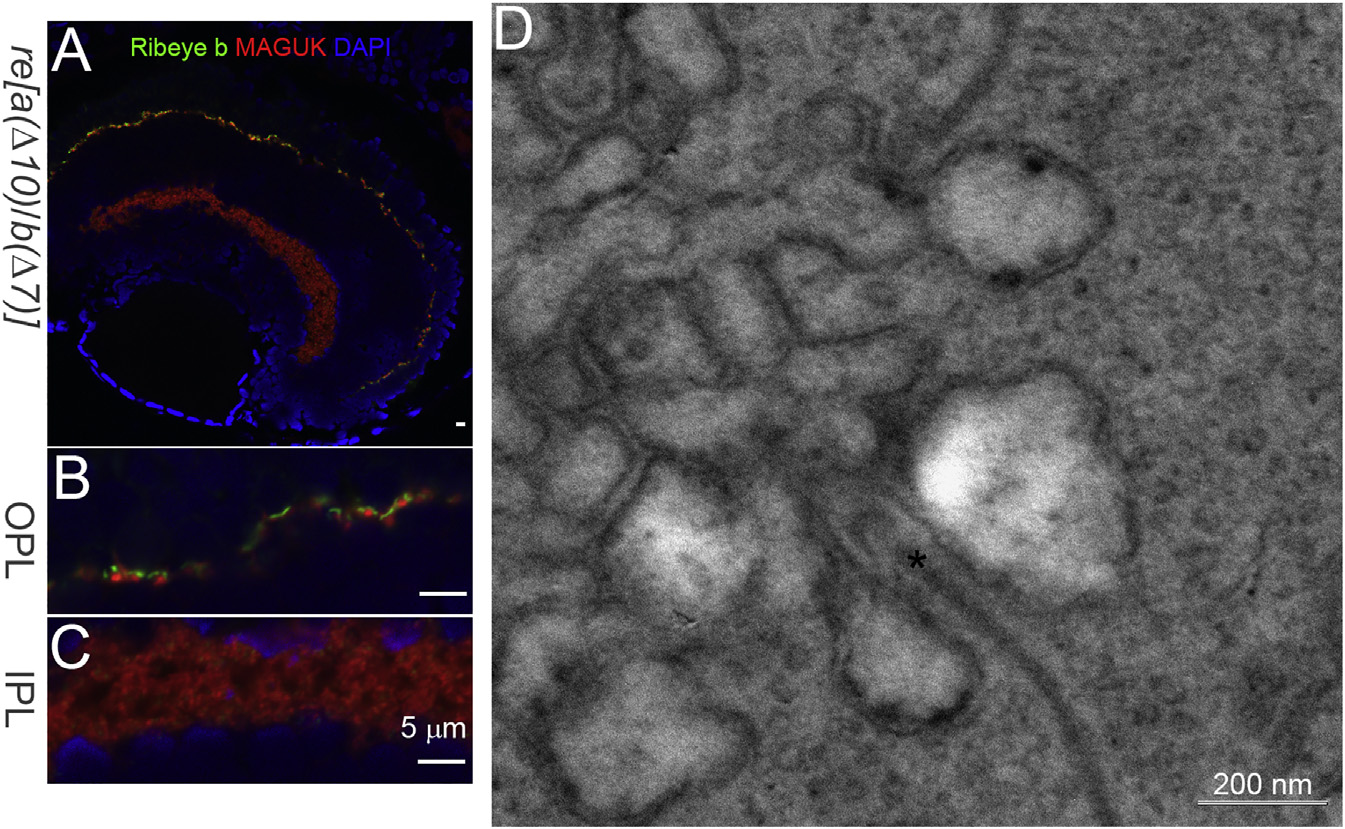
Fig. 1
Ribeye and Synaptic Ribbons Remain in the Retina of ribeye a(Δ10)/ ribeye b(Δ7) (re[a(Δ10)/b(Δ7)]) double homozygous mutants.
(A) Confocal image of Ribeye b (green) and post-synaptic maker MAGUK (red) staining in 5-dpf re[a(Δ10)/b(Δ7)] homozygous mutants. DAPI (blue) stains for the nucleus. Scale bar, 5 µm.
(B) 3× magnification of outer plexiform layer staining in 5-dpf re[a(Δ10)/b(Δ7)] homozygous mutants retina. Scale bar, 5 µm.
(C) 3× magnification of inner plexiform layer staining in 5-dpf re[a(Δ10)/b(Δ7)] homozygous mutants retina. Scale bar, 5 µm.
(D) Electron micrograph of photoreceptor ribbon from 5-dpf re[a(Δ10)/b(Δ7)] homozygous mutant retina. Scale bar, 200 nm.