
Fig. 3
- ID
- ZDB-FIG-160729-6
- Publication
- Lv et al., 2016 - Synaptic Ribbons Require Ribeye for Electron Density, Proper Synaptic Localization, and Recruitment of Calcium Channels
- Other Figures
- All Figure Page
- Back to All Figure Page
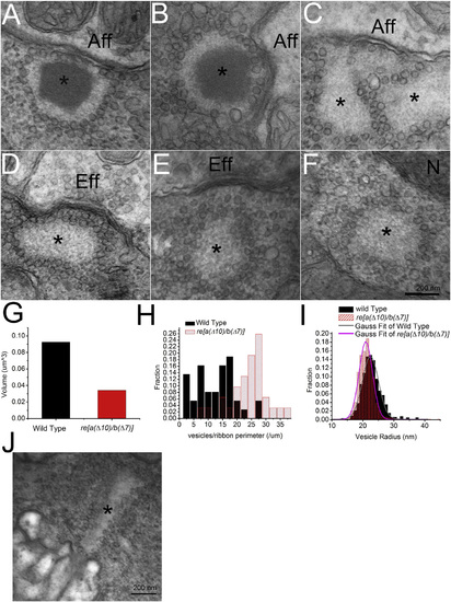
Mutant Zebrafish Exhibit Ribbons Lacking Synaptic Densities in Neuromast Hair Cells (A and B) Electron micrograph showing typical examples of synaptic ribbons in neuromast hair cells in 5- dpf WT zebrafish (marked by an asterisk). Note prominent density and typical localization near the afferent neuron (Aff). (C–F) Examples of electron micrographs of ghost ribbons (marked by an asterisk) taken from neuromast hair cells of re[a(Δ10)/b(Δ7)] homozygous zebrafish. Note the absence of a synaptic ribbon density with presence of vesicle array. Sone ghost ribbons locate near the afferent neurons (denoted Aff) (C), near the efferent neurons (Eff) (D), far away from the membrane (E), or in the middle of the cell (F). N denotes nucleus. Scale bar, 200 nm. (G) Volume of ribbon and ghost ribbons in 5-dpf WT and re[a(Δ10)/b(Δ7)] homozygous mutant hair cells. (H) Histogram of vesicle density per length of perimeter of ribbon or ghost ribbon in cross-section in WT and re[a(Δ10)/b(Δ7)] homozygous mutants. (I) Histogram of vesicle size associated with ribbon or ghost ribbon in WT and re[a(Δ10)/b(Δ7)] homozygous mutants. (J) Electron micrograph of ghost-ribbon like structure in the outer plexiform layer of re[a(Δ10)/b(Δ7)] double homozygous retina. |
| Fish: | |
|---|---|
| Observed In: | |
| Stage: | Day 5 |