
Fig. 4
- ID
- ZDB-FIG-160727-4
- Publication
- Liao et al., 2016 - Faster embryonic segmentation through elevated Delta-Notch signalling
- Other Figures
- All Figure Page
- Back to All Figure Page
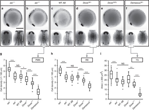
Elevated DeltaD increases the strength of lateral inhibition in the CNS. (a-f) Representative embryos stained with islet1, an early marker of primary neuronal differentiation. In each box, 4 somite-stage embryos lateral view, anterior to left (upper panel); dorsal view with anterior to top (lower left panel), of head showing bilateral trigeminal ganglia (TG); mid-trunk showing PMN at the midline, and RB neurons at lateral margin of neural plate (lower right panel). (a) Scale bars, 250µm. (g) Cell density of PMN. (h) Cell density of RB sensory neurons. (i) Area of the TG. Lower densities or smaller areas indicate higher Notch signalling strength. Data from aei-/- (n=33), aei+/- (n=18), WT (n=35), Dovertg/+ (n=14), Dovertg/tg (n=12) and Damascustg/+ (n≥2 independent trials. The central boxes of the box-and-whisker plots cover the interquartile range with median as line within box. Whiskers are 5th and 95th percentiles, extreme values are small bars. *P≤0.05, **P≤0.01, ***P≤0.001, NS, not significant (P>0.05), Student’s t-test. |