
Fig. S5
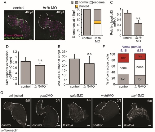
Characterisation of fn1bMO-injected embryos. (A) Immunofluorescence analysis shows reduced Fibronectin-positive staining (green) in the AVC of fli:nlsmCherry embryos injected with fn1bMO at 48 hpf (n=5 control, n=5 fn1bMO). (B) Quantification of overall morphological defects after fn1bMO injection (n=279 control, n=286 fn1bMO). (C) RT-qPCR analysis shows no significant increase in p53 expression following injection of fn1bbMO compared to controls (RNA extracted from 3 pools of 20 embryos). (D) Relative klf2a expression in klf2a:H2BEGFP embryos and (E) cell numbers at 48hpf are not affected by injection of fn1bMO (n=4 control, n=7 fn1bMO). (F) The flow profile was assessed by tracking red blood cells through the AVC in 3 consecutive heartbeats in control and fn1bMO embryos. The average proportion of forward (for), reversing (rev) and no flow (none) within the AVC is shown (n=2 and 3 embryos, respectively). The average maximum velocity (Vmax) in mm/s is also shown (blue text). (G) fli:kaede (control) and fli:klf2a embryos injected with gata2 and myh6 morpholinos were immunostained with antifibronectin antibody. Fibronectin synthesis can be seen throughout the heart in gata2 and myh6 morphants overexpressing klf2a. Error bar represents the standard deviation. Scale bars: 10 µm. |