
Fig. S3
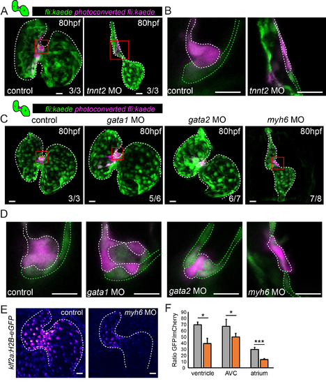
Defects in multi-layering occur in the presence of abnormal flow. Cells in the superior AVC of Tg(fli:kaede) embryos injected as shown were photoconverted in the superior AVC (schematic shown) at 48hpf and allowed to develop until 80hpf in all panels. (A) In tnnt2aMO-injected hearts, lacking heart contraction, multi-layering failed to occur by 80hpf (n=3 control, n=3 tnnt2aMO). Boxed red areas in (A) are shown in (B). Photoconverted cells remained in the single-layered endocardium. White dashed line highlights the photoconverted cells of the superior AVC. (C) Many cells were observed in the cardiac jelly of gata1MO-injected hearts (yellow dashed line highlights cells in the cardiac jelly) and intermingling of non-photoconverted (green) and photoconverted (magenta) cells was sometimes observed (n=3/6) (D). Boxed red areas from (C) are shown in (D). In contrast, multi-layering was impaired in gata2MO-injected and failed to occur in myh6 MOinjected embryos. n=3 control, n=6 gata1MO, n=7 gata2MO, n=8 myh6 MO. Green dashed line highlights the non-photoconverted cells of the superior AVC. (E and F) Quantification of GFP signal intensity in klf2a:H2B-eGFP embryos demonstrates reduced klf2a expression in myh6 MO hearts (n=4 control hearts, n=3 myh6MO hearts). Error bar represents the standard deviation. Scale bars: 10 µm. |