
Fig. 5
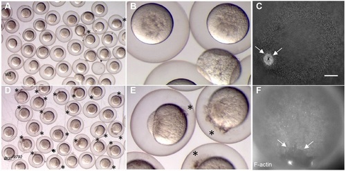
Wound healing defect in aura mutant embryos. (A,B,D,E) Embryos from wild-type (A,B) and aura mutant (D,E) mothers were pricked with a glass injection needle at 10-15mpf and allowed to recover until 30mpf. After recovery, most wild-type embryos have resealed their membrane, whereas a majority of aura mutant embryos show continued leakage of yolk and ooplasm (D,E, asterisks). (C,F) Wild-type (C) and aura mutant (F) embryos were fixed 1min after wounding and labeled with phalloidin. Wild-type embryos recruit F-actin to the closing wound edge (C, arrows; 7/7). All aura embryos examined show reduced F-actin enrichment at the wound edge and a larger wound diameter (F, arrows; 13/13). Scale bar: 100µm in C,F. |
| Fish: | |
|---|---|
| Condition: | |
| Observed In: | |
| Stage: | 1-cell |