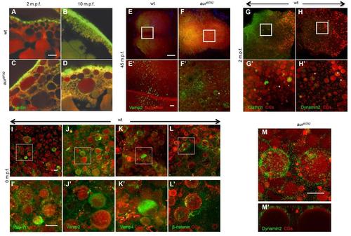
Membrane dynamics of wild-type and aura mutant embryos. (A-D) Side view of wild-type (A,B) and aura mutant (C,D) embryos at 2 mpf and 10 mpf, labeled to detect F-actin and CGs, showing that mutants exhibits a similar localization of CGs at the cortex during egg activation (A,C) and retains a fraction of CGs (B,D). (E-F) In wild-type at 45 mpf, β-catenin localizes to the furrow but Vamp2 does not yet localize to this structure (4/4 embryos) (E, E′) In aura mutants, Vamp2 appears localize to ectopic CGs (8/9 embryos) (F,F′). Vamp2 also localizes to small cortical particles in both wild-type and mutant embryos (E′, F′). (G-L) Localization of Clathrin (G, G′), Dynamin2 (H,H′), Rab11 (I,I′), Vamp2 (J,J′), Vamp4 (K,K′), and ß-catenin (L,L′) to a subset of CGs in wild-type at 2 mpf (G-H) or 0 mpf (I-L). (M, M′) Dynamin2 surrounding CGs in aura mutant embryos as seen by a face-on view (M) and a reconstructed orthogonal view (M′). Scale bars: A-D (bar in A): 10 µm; E-H (bar in E): 100 µm; E′-H′(bar in E′): 10 µm; I-M′ (bar in I,I′,M): 10 µm.
|
