|
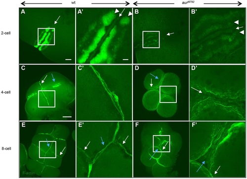
aura mutant embryos exhibit reduced accumulation of pericleavage and adhesive junction F-actin. (A,A′) In wild type (25/25), F-actin becomes recruited to the contractile ring (arrows) and pericleavage regions (arrowheads). (B,B′) In aura mutants, the contractile ring pericleavage F-actin is reduced (30/30). (C,C′) At the 4-cell stage, the furrow corresponding to the first cell cycle has formed adhesion junctions containing F-actin (white arrow). (D,D′) At the same stage, aura embryos do not exhibit adhesion junction F-actin cables (white arrow). (E-F′) Defects continue to be observed in subsequent furrows. White and blue arrows indicate furrows for the first and second cell cycles, respectively. (A′-F′) Higher magnification views of boxed regions in A-F. Scale bars: 20µm in A,B; 100µm in C-F; 10µm in A′-F′.
|