
Fig. 4
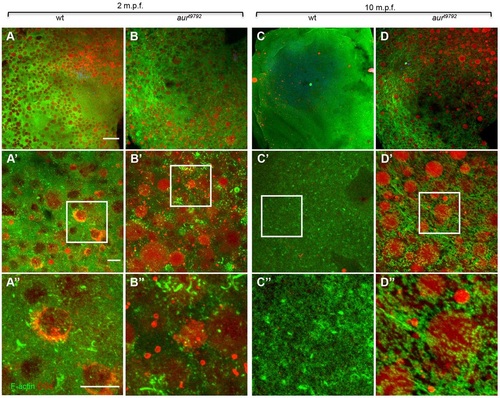
aura mutants retain CGs. (A,B) During egg activation (2mpf), CGs appear embedded in an F-actin network in both wild type (A) and aura mutants (B). (C,D) By 10mpf, wild-type eggs have extruded nearly all CGs (C), whereas a large fraction of CGs are retained in aura mutants (D) (wild type, 0 mpf n=152, 10 mpf n=3; mutant, 0 mpf n=245, 10 mpf n=130; chi-square, P<0.0001). By 10mpf, the cortical F-actin cytoskeleton appears as a network of short F-actin fibers in aura mutants (D′′), in contrast to being largely disassembled as in wild type (C′′). (A′-D′) Higher magnification views of A-D; the boxed regions are further magnified in A′′-D′′. Scale bars: 100µm in A-D; 10µm in A′-D′,A′′-D′′. |
| Fish: | |
|---|---|
| Observed In: | |
| Stage: | 1-cell |