
Fig. 7
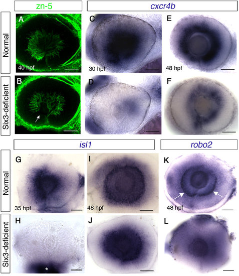
Delayed differentiation of RGCs in Six3-deficient retinas. (A-L) Eyes in whole embryos (A-F) or dissected eyes (G-L). (A,B) zn-5 immunohistochemistry of eyes in normal (A) and Six3-deficient embryos (B). Arrow in (B) points at the anterior-ventral region lacking differentiated RGCs. (C-F) WISH for cxcr4b at 30 hpf (C,D) and 48 hpf (E,F) in normal (C,E) and Six3-deficient (D,F) retinas. (G-J) WISH for isl1 at 35 hpf (G,H) and 48 hpf (I,J) in normal (G,I) and Six3-deficient (H,J) retinas. Asterisk in H marks remnants of tissues outside the eye, which was dissected. (K-L) WISH for robo2 at 48 hpf in normal (K) and Six3-deficient (L) retinas. Arrows in (K,L) point at RGCs that express robo2. All panels are lateral views, anterior to the left. Scale bars are 50 µm. |
| Genes: | |
|---|---|
| Antibody: | |
| Fish: | |
| Anatomical Terms: | |
| Stage Range: | Prim-15 to Long-pec |
| Fish: | |
|---|---|
| Observed In: | |
| Stage Range: | Prim-15 to Long-pec |