
Fig. 3
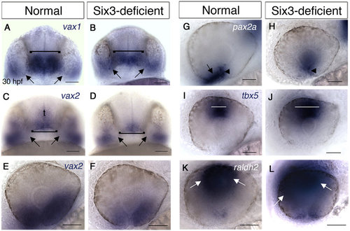
Eyes and optic stalks of Six3-deficient embryos are abnormally patterned. (A-L) WISH of 30 hpf normal (A,C,E,G,I,K) and Six3-deficient (B,D,F,H,J,L) embryos. (A,B) vax1 expression. Brackets marks POAs, arrows point at OSs. (C-F) vax2 expression in the POA, OS and ventral retina. Brackets marks POAs, arrows point at OSs. t, telencephalon. (G,H) pax2a expression in the optic fissure (arrowheads in G,H) and OS (arrow in G, missing in H). (I,J) tbx5 expression in dorsal retina. White lines mark the width of the region where high tbx5 levels are observed. (K,L) raldh2 expression in the dorsal retina. Arrows mark limits of high raldh2 expression. (A-D) are ventral views, anterior is up. E-L are lateral views, anterior to the left, dorsal up. Scale bars are 50 µm. |
| Genes: | |
|---|---|
| Fish: | |
| Anatomical Terms: | |
| Stage: | Prim-15 |
| Fish: | |
|---|---|
| Observed In: | |
| Stage: | Prim-15 |